
Datoteka:1gwe antipar betaSheet both.png

Veličina ovog prikaza: 800 × 440 piksela. Ostale rezolucije: 320 × 176 piksela | 640 × 352 piksela | 1.024 × 563 piksela | 1.280 × 704 piksela | 2.000 × 1.100 piksela.
Izvorna datoteka (2.000 × 1.100 piksela, veličina datoteke: 1,04 MB, MIME tip: image/png)
| Ova datoteka je s Wikimedia Commonsa. Opis s njene stranice opisa datoteke prikazan je ispod. Commons je skladište slobodnih medija i datoteka za sve projekte fondacije Wikimedia. Možete i Vi pomoći. |
Sažetak
| Opis |
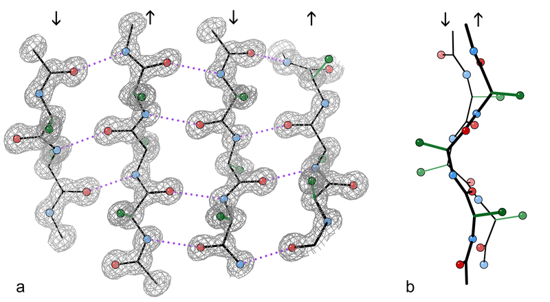
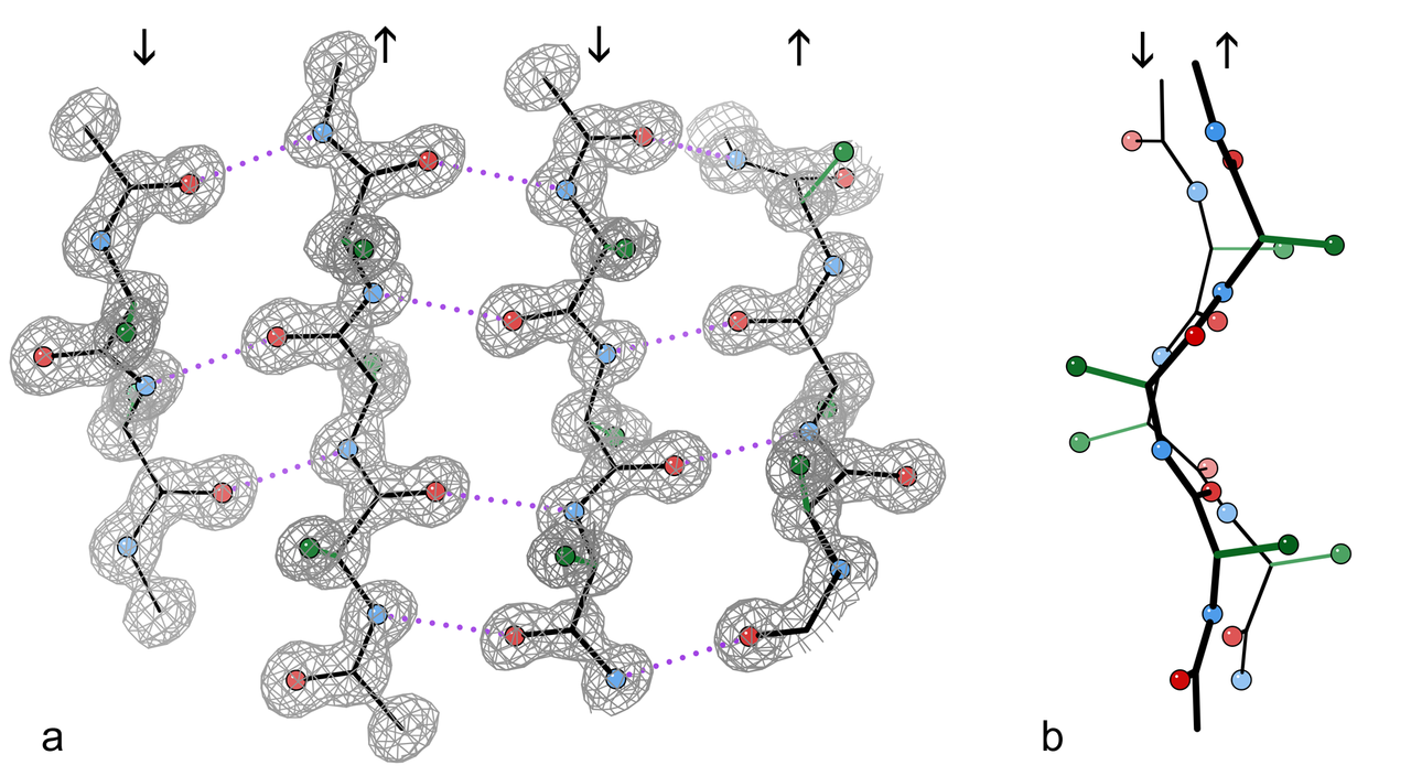
English: An example of a 4-stranded antiparallel β sheet fragment from a crystal structure of the enzyme catalase (PDB file 1GWE at 0.88 Å resolution). a) Front view, showing the antiparallel hydrogen bonds (dotted) between peptide NH and CO groups on adjacent strands. Arrows indicate chain direction, and electron density contours outline the non-H atoms. O atoms are red balls, N atoms are blue, and H atoms are omitted for simplicity; sidechains are shown only out to the first sidechain C atom (green). b) Edge-on view of the central two β strands in a, showing the righthanded twist and the pleat of Cαs and sidechains that alternately stick out in opposite directions from the sheet.
Français : Exemple d'un fragment de feuillet β à quatre chaines antiparallèles extrait de la structure cristalline de l'enzyme catalase (résolution 0,88 Å). a) Vue de face, montrant les liaisons hydrogènes (en pointillés) entre les groupes NH et CO des acides aminés adjacents. Les flèches indiquent l'orientation des chaines, et les contours de densité d'électron entourent les atomes autres que l'hydrogène. Les atomes d'oxygène sont donnés en rouge, ceux d'azote en bleu. Les atomes d'hydrogène sont omis pour plus de simplicité. Dans le même but, seul le premier carbone des radicaux est montré (en vert). b)vue par côté des deux chaines centrales montrant la torsion à droite des chaines l'une par rapport à l'autre, ainsi que les plis de chacune d'elle qui orientent les carbones portant les radicaux des acides aminés alternativement de part et d'autre de celles-ci. |
| Datum | |
| Izvor | Vlastito djelo |
| Autor | Dcrjsr |
Licenciranje
Ja, vlasnik autorskog prava ovog djela, ovdje ga objavljujem pod sljedećom licencom:
Ova datoteka je licencirana pod Creative Commons Attribution 3.0 neportiranom licencom.
- Slobodni ste:
- da dijelite – da kopirate, distributirate i prenosite djelo
- da remiksate – da prilagodite djelo
- Pod sljedećim uslovima:
- pripisivanje – Morate pripisati odgovarajuće autorske zasluge, osigurati link ka licenci i naznačiti jesu li napravljene izmjene. To možete uraditi na bilo koji razumni način, ali ne tako da se sugerira da davalac licence odobrava Vas ili Vašu upotrebu njegovog djela.

|
This image has been assessed under the valued image criteria and is considered the most valued image on Commons within the scope: Protein sheets and strands. You can see its nomination here. |
Historija datoteke
Kliknite na datum/vrijeme da vidite verziju datoteke iz tog vremena.
| Datum/vrijeme | Smanjeni pregled | Dimenzije | Korisnik | Komentar | |
|---|---|---|---|---|---|
| trenutno | 18:00, 10 april 2010 | | 2.000 × 1.100 (1,04 MB) | Dcrjsr | {{Information |Description={{en|1=An example of a 4-stranded antiparallel β sheet fragment from a crystal structure of the enzyme catalase (PDB file 1GWE at 0.88Å resolution). a) Front view, showing the antiparallel hydrogen bonds (dotted) between pepti |
Upotreba datoteke
Sljedeća stranica koristi ovu datoteku:
Globalna upotreba datoteke
Sljedeći wikiji koriste ovu datoteku:
- Upotreba na ar.wikipedia.org
- Upotreba na bg.wikipedia.org
- Upotreba na ca.wikipedia.org
- Upotreba na en.wikipedia.org
- Upotreba na en.wikibooks.org
- Upotreba na fa.wikipedia.org
- Upotreba na fr.wikipedia.org
- Upotreba na gl.wikipedia.org
- Upotreba na ja.wikipedia.org
- Upotreba na mk.wikipedia.org
- Upotreba na pl.wikipedia.org
- Upotreba na ru.wikipedia.org
- Upotreba na sh.wikipedia.org
- Upotreba na sr.wikipedia.org
- Upotreba na tr.wikipedia.org