
Datoteka:Fundus of patient with retinitis pigmentosa, mid stage.jpg
Veličina ovog prikaza: 699 × 599 piksela. Ostale rezolucije: 280 × 240 piksela | 560 × 480 piksela | 871 × 747 piksela.
Izvorna datoteka (871 × 747 piksela, veličina datoteke: 107 KB, MIME tip: image/jpeg)
| Ova datoteka je s Wikimedia Commonsa. Opis s njene stranice opisa datoteke prikazan je ispod. Commons je skladište slobodnih medija i datoteka za sve projekte fondacije Wikimedia. Možete i Vi pomoći. |
| Opis |
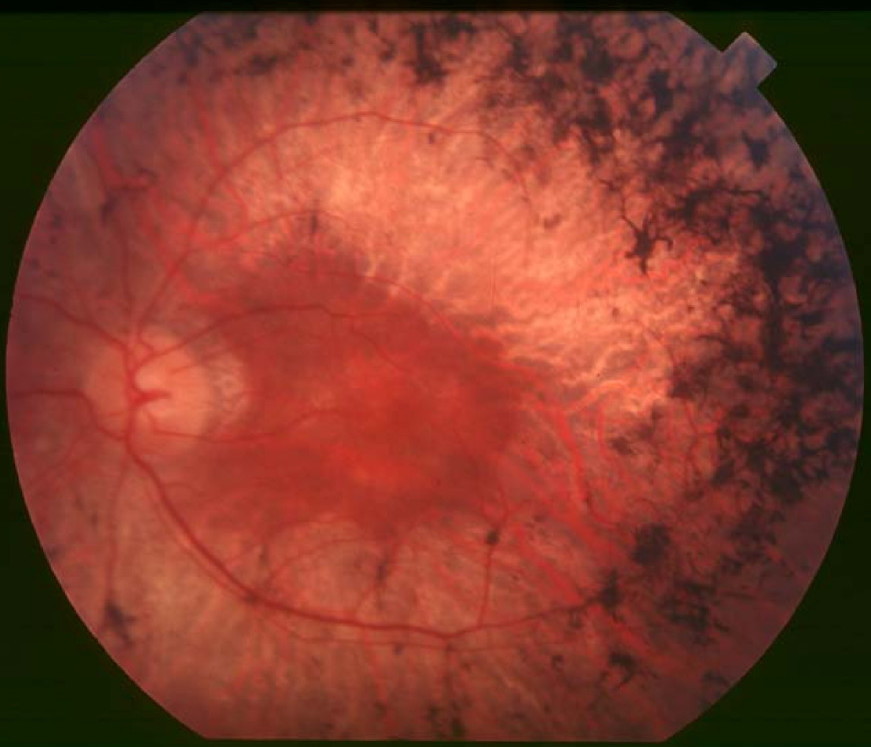
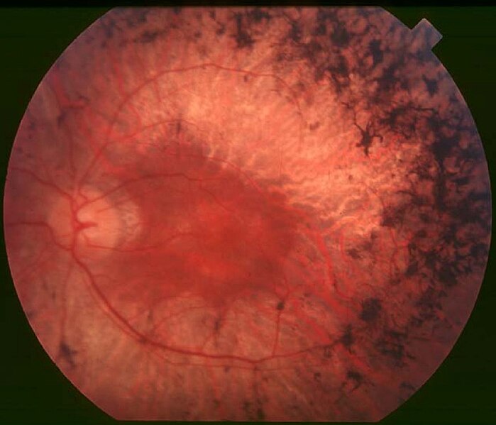
English: Figure 2. Fundus of patient with retinitis pigmentosa, mid stage (Bone spicule-shaped pigment deposits are present in the mid periphery along with retinal atrophy, while the macula is preserved although with a peripheral ring of depigmentation. Retinal vessels are attenuated.) Hamel Orphanet Journal of Rare Diseases 2006 1:40 doi:10.1186/1750-1172-1-40 |
| Datum | |
| Izvor | Retinitis pigmentosa by Christian Hamel |
| Autor | Christian Hamel |
| Dopuštenje (Naknadno korištenje ove datoteke) |
© 2006 Hamel; licensee BioMed Central Ltd. This is an Open Access article distributed under the terms of the Creative Commons Attribution License (https://creativecommons.org/licenses/by/2.0), which permits unrestricted use, distribution, and reproduction in any medium, provided the original work is properly cited. |
Ova datoteka je licencirana pod Creative Commons Attribution 2.0 generičkoj licenci.
- Slobodni ste:
- da dijelite – da kopirate, distributirate i prenosite djelo
- da remiksate – da prilagodite djelo
- Pod sljedećim uslovima:
- pripisivanje – Morate pripisati odgovarajuće autorske zasluge, osigurati link ka licenci i naznačiti jesu li napravljene izmjene. To možete uraditi na bilo koji razumni način, ali ne tako da se sugerira da davalac licence odobrava Vas ili Vašu upotrebu njegovog djela.
Historija datoteke
Kliknite na datum/vrijeme da vidite verziju datoteke iz tog vremena.
| Datum/vrijeme | Smanjeni pregled | Dimenzije | Korisnik | Komentar | |
|---|---|---|---|---|---|
| trenutno | 12:17, 2 decembar 2017 | | 871 × 747 (107 KB) | Doc James | Cropped 27 % horizontally and 7 % vertically using CropTool with precise mode. |
| 15:52, 22 septembar 2009 |  | 1.200 × 799 (126 KB) | CopperKettle | {{Information |Description={{en|1=Figure 2. Fundus of patient with retinitis pigmentosa, mid stage (Bone spicule-shaped pigment deposits are present in the mid periphery along with retinal atrophy, while the macula is preserved although with a peripheral |
Upotreba datoteke
Sljedeća stranica koristi ovu datoteku:
Globalna upotreba datoteke
Sljedeći wikiji koriste ovu datoteku:
- Upotreba na ar.wikipedia.org
- Upotreba na ca.wikipedia.org
- Upotreba na da.wikipedia.org
- Upotreba na en.wikipedia.org
- Upotreba na en.wikiversity.org
- Upotreba na es.wikipedia.org
- Upotreba na eu.wikipedia.org
- Upotreba na fa.wikipedia.org
- Upotreba na fi.wikipedia.org
- Upotreba na fr.wikipedia.org
- Upotreba na he.wikipedia.org
- Upotreba na hy.wikipedia.org
- Upotreba na it.wikipedia.org
- Upotreba na ko.wikipedia.org
- Upotreba na la.wikipedia.org
- Upotreba na or.wikipedia.org
- Upotreba na outreach.wikimedia.org
- Upotreba na pl.wikipedia.org
- Upotreba na pt.wikipedia.org
- Upotreba na ru.wikipedia.org
- Upotreba na sl.wikipedia.org
- Upotreba na sr.wikipedia.org
- Upotreba na sv.wikipedia.org
- Upotreba na th.wikipedia.org
- Upotreba na tr.wikipedia.org
- Upotreba na tt.wikipedia.org
- Upotreba na uk.wikipedia.org
- Upotreba na vi.wikipedia.org
Pogledajte globalne upotrebe ove datoteke.