Datoteka:Diseases of the nervous system (1910) (14586639887).jpg

Sadržaj stranice nije podržan na drugim jezicima.
S Wikipedije, slobodne enciklopedije

Izvorna datoteka(2.864 × 1.892 piksela, veličina datoteke: 726 KB, MIME tip: image/jpeg)

Sažetak

Opis
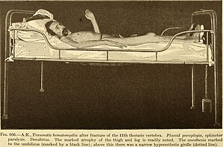
English:

Identifier: diseasesofnervou00chur (find matches)
Title: Diseases of the nervous system
Year: 1910 (1910s)
Authors: Church, Archibald, b. 1861, ed Salinger, Julius L. (Julius Lincoln), tr
Subjects: Nervous system
Publisher: New York and London : D. Appleton and company
Contributing Library: The Library of Congress
Digitizing Sponsor: The Library of Congress

View Book Page: Book Viewer
About This Book: Catalog Entry
View All Images: All Images From Book
Click here to view book online to see this illustration in context in a browseable online version of this book.

Text Appearing Before Image:
rtion of the cord was contracted, and stained a uniform light yellow.In the higher sections (Fig. 167 a-e) up into the cervical cord the usual picture ofascending degeneration in the posterior columns as well as in the periphery of thelateral columns was recognized and could be noted by the paler stain. Besides a focusof softening about the size of a millet seed (Fig. 167 e) was found from the 8th dorsalsegment, extending downward in the left posterior column near the commissure beingdistributed to the dorsal cord, and at the 4th dorsal segment widening into a cavity 400 MYELITIS (167 d) thence extending over the middle of both posterior columns and filled withyellowish masses. This tubular focus terminated in the lower cervical cord. Leyden, upon histologic examination (according to Nissl, Marchi, Weigert, etc.),was unable to find nerve fibers or ganglion cells in the lower dorsal, lumbar or sacralcord; the tissue in the entire transverse section consisted of numerous granular cells,
Text Appearing After Image:
proliferated glia meshes, myelin granules and a few hematoidin crystals. Only in afew longitudinal sections were there scant fragments of destroyed axis cylinders andmedullary sheaths. The pia mater was greatly contracted, the posterior roots wereonly in part destroyed, while the anterior roots were transformed into an almost com-plete dense, fibrillary tissue. This destruction of the spinal cord continued in thelateral columns to the 8th dorsal segment, while in the posterior columns it could befollowed, in the form of a peg-like focus to the lower cervical cord. The largest focus THE CLINICAL PICTURE OF DORSAL MYELITIS 401 also situated in the 4th dorsal segment (Fig. 167 d) consists of granular cells, remainsof medullary sheaths and glia fibers, the latter becoming denser around the focus.With this there is the usual ascending degeneration in the posterior columns (Fig.167 ci-e) and in the lower border zone (cerebellar lateral column, Gowers bundle).The muscles of the leg were in a

Note About Images

Please note that these images are extracted from scanned page images that may have been digitally enhanced for readability - coloration and appearance of these illustrations may not perfectly resemble the original work.
Datum
Izvor

https://www.flickr.com/photos/internetarchivebookimages/14586639887/

Autor

Church, Archibald, b. 1861, ed;

Salinger, Julius L. (Julius Lincoln), tr
Dopuštenje
(Naknadno korištenje ove datoteke)
At the time of upload, the image license was automatically confirmed using the Flickr API. For more information see Flickr API detail.
Ostale verzije
Flickr tags
InfoField
Flickr posted date
InfoField
29 juli 2014


Licenciranje

This image was taken from Flickr's The Commons. The uploading organization may have various reasons for determining that no known copyright restrictions exist, such as:
  1. The copyright is in the public domain because it has expired;
  2. The copyright was injected into the public domain for other reasons, such as failure to adhere to required formalities or conditions;
  3. The institution owns the copyright but is not interested in exercising control; or
  4. The institution has legal rights sufficient to authorize others to use the work without restrictions.

More information can be found at https://flickr.com/commons/usage/.


Please add additional copyright tags to this image if more specific information about copyright status can be determined. See Commons:Licensing for more information.
This image was originally posted to Flickr by Internet Archive Book Images at https://flickr.com/photos/126377022@N07/14586639887. It was reviewed on 27 septembar 2015 by FlickreviewR and was confirmed to be licensed under the terms of the No known copyright restrictions.

27 septembar 2015

Captions

Add a one-line explanation of what this file represents

Items portrayed in this file

prikazuje

MIME type engleski

image/jpeg

Historija datoteke

Kliknite na datum/vrijeme da vidite verziju datoteke iz tog vremena.

Datum/vrijemeSmanjeni pregledDimenzijeKorisnikKomentar
trenutno02:01, 19 oktobar 2016Smanjeni pregled verzije na dan 02:01, 19 oktobar 20162.864 × 1.892 (726 KB)SteinsplitterBotBot: Image rotated by 90°
09:30, 27 septembar 2015Smanjeni pregled verzije na dan 09:30, 27 septembar 20151.892 × 2.868 (729 KB)== {{int:filedesc}} == {{information |description={{en|1=<br> '''Identifier''': diseasesofnervou00chur ([https://commons.wikimedia.org/w/index.php?title=Special%3ASearch&profile=default&fulltext=Search&search=insource%3A%2Fdiseasesofnervou00chur%2F fin...

Sljedeća stranica koristi ovu datoteku:

Globalna upotreba datoteke

Sljedeći wikiji koriste ovu datoteku: